
HEMMAGINERO project ID
| Title: | Hemoglobin-Based Spectroscopy and Nonlinear Imaging of Erythrocytes and Their Membranes as Emerging Diagnostic Tool |
| Acronym: | HEMMAGINERO |
| Funding agency: | Science Fund of Republic of Serbia |
| Call (deadline): | PROMIS (02.09.2019.) |
| Start date: | 01.09.2020. |
| Duration: | 2 years |
| Title in Serbian: | Спектроскопија и нелинеарно осликавање еритроцита и њихових мембрана засновани на хемоглобину као полазница за развој нове дијагностичке методе |
HEMMAGINERO is one of 59 funded projects out of 585 applications in the PROMIS call.

The team

|
Institutions
Facebook page (news and gallery)
https://www.facebook.com/Hemmaginero
HEMMAGINERO in brief
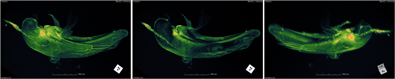
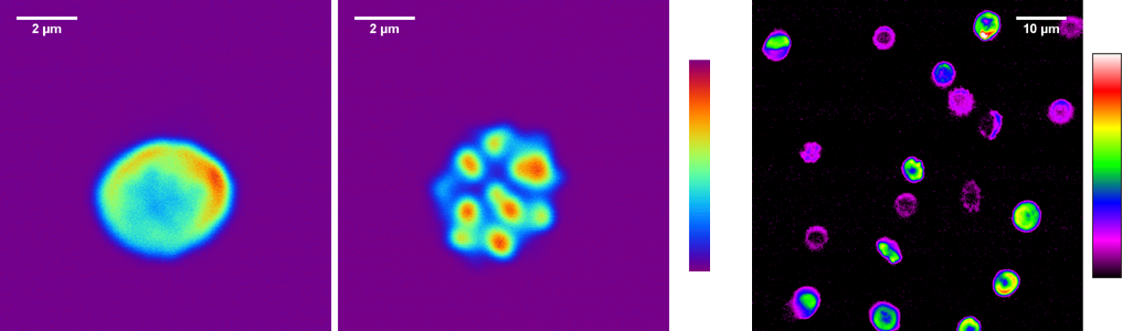
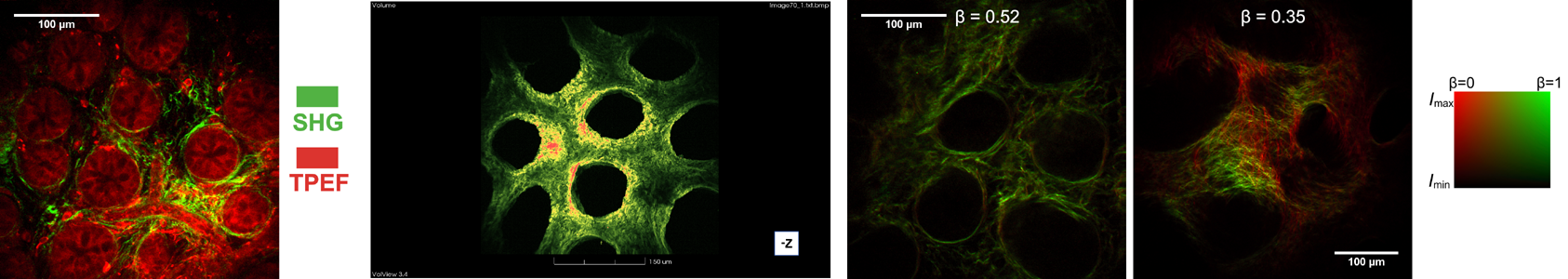
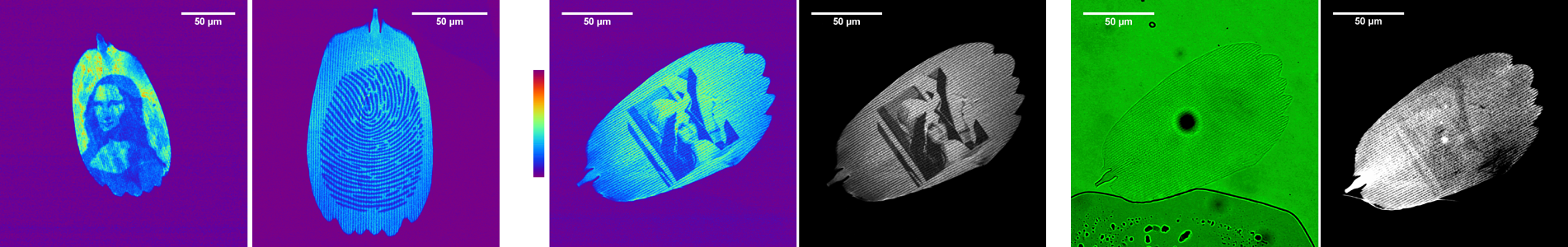
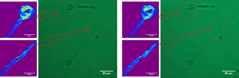
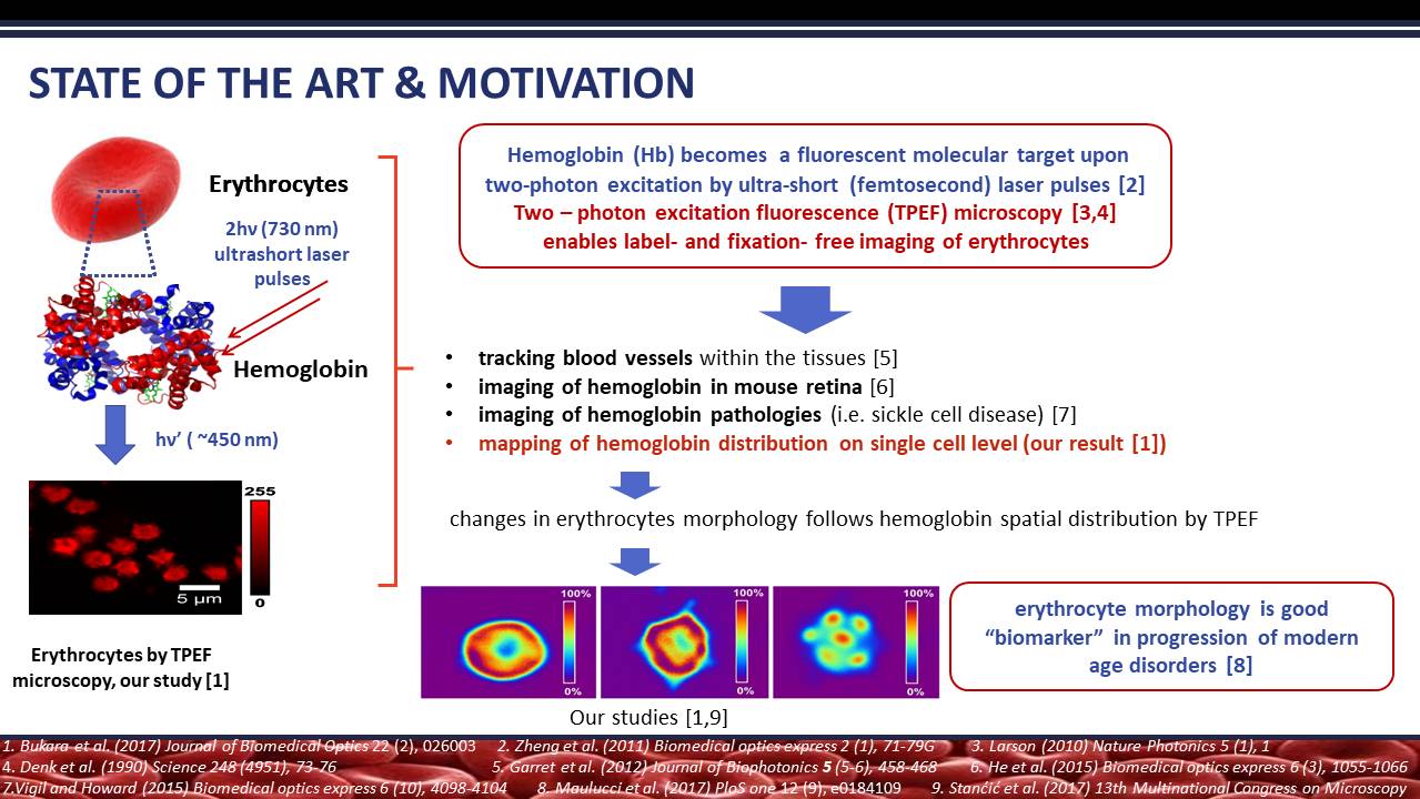
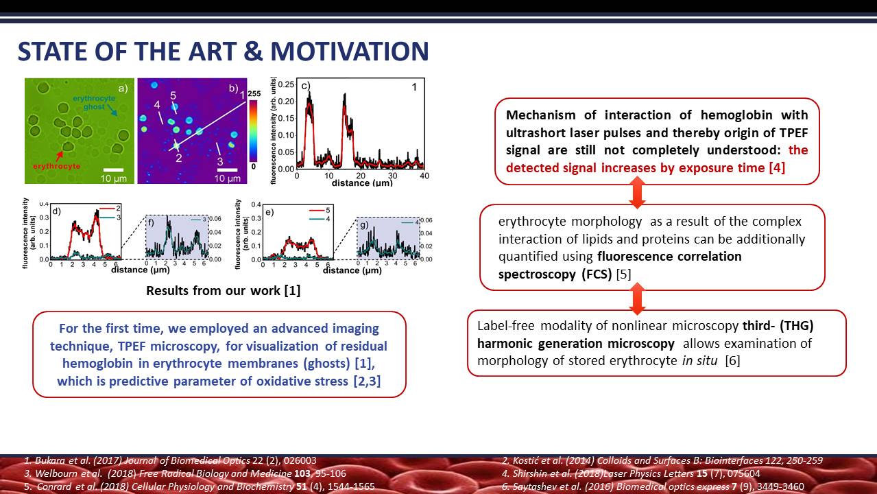
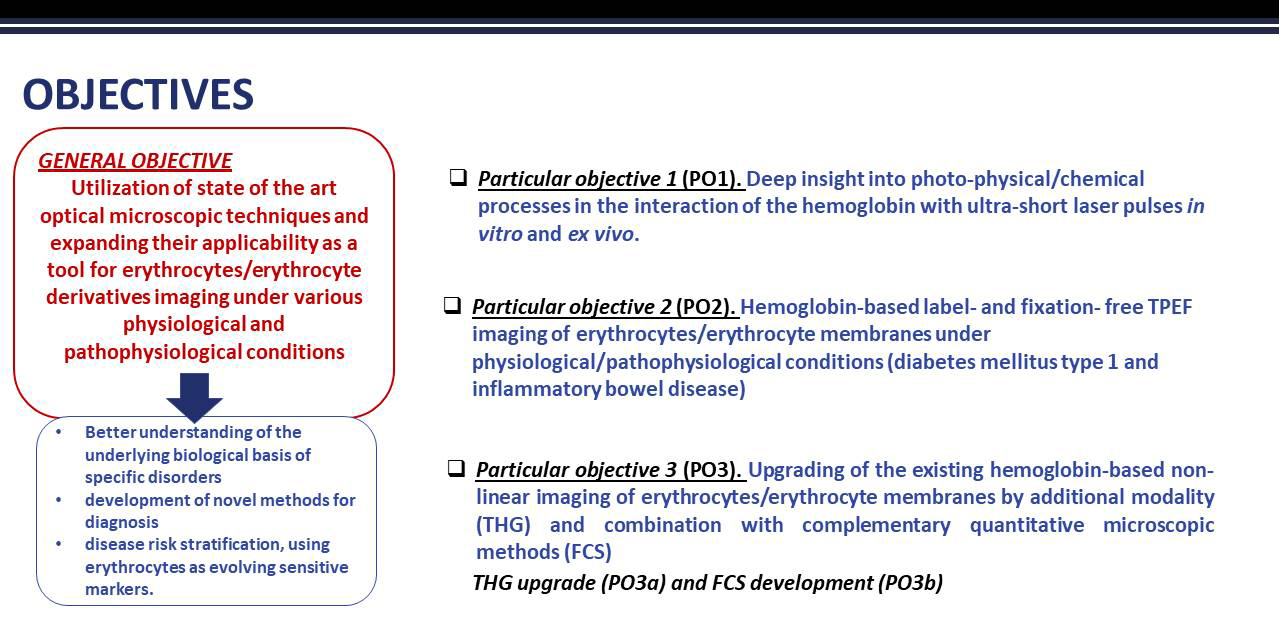
In the HEMMAGINERO project, we are aiming to utilize state-of-the art optical microscopic techniques expanding their applicability for erythrocytes and erythrocyte derivatives imaging as a potential diagnostic method for modern age disorders, such as cancer, diabetes and other inflammatory/autoimmune diseases. Our research will be mostly based on two-photon excitation fluorescence microscopy (TPEF) and recently demonstrated intrinsic feature of hemoglobin to become a fluorescent molecular target upon two-photon excitation by ultra-short laser pulses. We will unite our "know-how" on TPEF with third harmonic generation (THG) imaging of erythrocytes and complement it with quantitative information obtained by fluorescence correlation spectroscopy (FCS). The latter two will be developed within this project. Using erythrocytes as sensitive markers, we will demonstrate how combining the data acquired by custom-made techniques and established protocols for in vitro and ex vivo hemoglobin based imaging can contribute to a better understanding of the underlying biological basis of specific disorders. This is the first step in development of novel methods for diagnosis and disease risk stratification. We will also boost our initiated research on TPEF microscopy as an effective tool for label- and fixation -free imaging of erythrocytes and their membranes at single cell level. The research revealed the relation between spatial distribution of hemoglobin within erythrocytes and their morphology implying that this relation is an indicator of erythrocytes functional status.
Six researchers from two leading national institutions and from various, complementary, fields (physics, biology, biophotonics, pharmacy) will carry out the activities divided into 7 tasks and deliver 13 items, amongst two new techniques will be developed: THG and FCS for the benefit to the broad international and Serbian scientific community. HEMMAGINERO is supported by DESY, Germany and Karolinska Institutet, Sweden formally and by open access to the equipment. The project will be realized in 2 years and by cca 200,000 EUR employing and upgrading the most valuable equipment in Serbian scientific institutions and respecting all the safety and ethical issues.
Motivation
The motivation for the proposed project is to boost our already initiated research, which demonstrated that in-house TPEF microscopy set-up is effective tool for label- and fixation -free imaging of erythrocytes and their membranes, so called ghosts, at single cell level [1]. For the first time by employing TPEF microscopy we revealed the relation between spatial distribution of hemoglobin within erythrocytes and their morphology [1], which is known to be indicator of erythrocytes functional status [2]. Based on our pilot investigation, we are inspired to propose the research on further development of non-linear imaging modalities for erythrocytes/erythrocyte derivatives in various physiological and pathophysiological states. This would enable us to gather more detailed information on sub-micron scales about the finely tuned bidirectional interaction of these entities with physiological/pathophysiological environment, relevant for diagnostic purposes. The idea is to contribute to the understanding of the photo-physical/chemical processes underlying the mentioned interaction of hemoglobin with ultra-short laser pulses, what will lead to more efficient implementation of developed techniques in real settings. Additional motivational force is that proposed research project, will generate the new multidisciplinary team of young scientists in the Republic of Serbia, launching research in advanced imaging and microscopic techniques TPEF, THG and FCS, which would be on disposal for the interdisciplinary research in relevant local institutions as well.
[1] Bukara, K. et al., Mapping of hemoglobin in erythrocytes and erythrocyte ghosts using two photon excitation fluorescence microscopy. Journal of Biomedical Optics 22 (2), 026003 (2017).
[2] Tsakanova, G. et al., Two-photon microscopy imaging of oxidative stress in human living erythrocytes. Biomedical Optics Express 8 (12), 5834-5846 (2017).
Hemoglobin and erythrocytes imaging by TPEF
Objectives (goals)
Project scheme

Presentation of the project in PDF